Why a Physician Might Order a Sleep Study
Persistent fatigue despite adequate time in bed, a partner reporting loud snoring or witnessed pauses in breathing, unexplained morning headaches, or episodes of acting out dreams — these are the kinds of symptoms that prompt a sleep medicine referral. A sleep study gives clinicians objective, sensor-derived evidence of what is actually happening while a patient sleeps, something no amount of self-reported history can fully replicate.
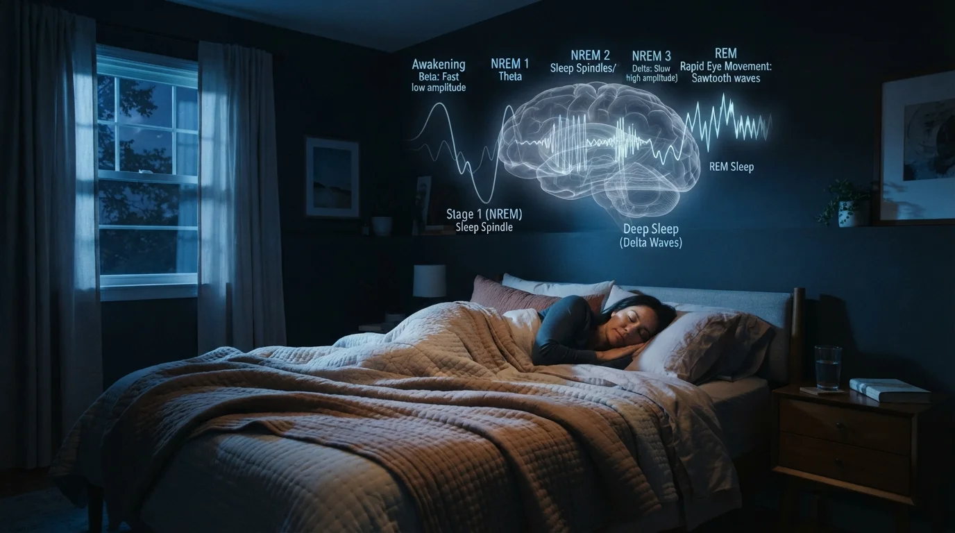
Sleep disorders affect tens of millions of Americans, yet many go undiagnosed for years because symptoms overlap with ordinary tiredness or stress. Polysomnography creates a detailed, time-stamped record that allows specialists to distinguish between conditions — for instance, separating obstructive sleep apnea (where the airway physically collapses) from central sleep apnea (where the brain momentarily stops signaling the respiratory muscles). Understanding how sleep stages are structured helps explain why disruptions at different points in the night produce very different symptoms.
What Happens Before You Go to Sleep
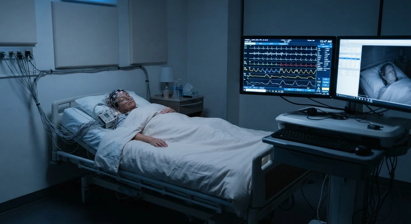
When you arrive at the sleep lab, a technologist will walk you through the process and answer questions. The setup phase — attaching sensors — takes roughly 30 to 60 minutes. Here is what is typically applied:
- EEG electrodes (electroencephalography): Small metal discs attached to the scalp with conductive paste to record brain wave activity and identify sleep stages.
- EOG electrodes (electrooculography): Placed near the outer corners of the eyes to track eye movements, particularly the rapid eye movements that define REM sleep.
- EMG electrodes (electromyography): Applied to the chin and legs to detect muscle tone and movement, which is relevant for diagnosing conditions such as REM sleep behavior disorder and periodic limb movement disorder.
- Respiratory sensors: Belts around the chest and abdomen measure breathing effort; a cannula near the nostrils records airflow.
- Pulse oximeter: A clip on the fingertip continuously measures blood oxygen saturation.
- ECG leads: Monitor heart rate and rhythm throughout the night.
The wires connect to a lightweight junction box that allows you to move relatively freely, including walking to the bathroom. None of the sensors pierce the skin.
~30 million
Americans estimated to have obstructive sleep apnea
According to the American Academy of Sleep Medicine, a large proportion of OSA cases remain undiagnosed in the U.S.
80%+
Estimated proportion of moderate-to-severe OSA cases undiagnosed
Research published in sleep medicine literature consistently finds that the majority of people with clinically significant sleep apnea have not received a formal diagnosis.
7–8 channels
Minimum simultaneous signal streams in a standard sleep study
Full polysomnography records brain waves, eye movements, muscle activity, heart rhythm, airflow, respiratory effort, and blood oxygen simultaneously.
What Clinicians Are Actually Measuring
During the study, a technologist monitors the incoming data streams from an adjacent room and can communicate with you through an intercom if needed. The recording captures an enormous amount of information:
Sleep architecture — the pattern and proportion of NREM stages (N1, N2, N3) and REM sleep across the night. Abnormal architecture can signal a range of disorders. For deeper context on what these stages do for your brain and body, see our article on what the brain does during sleep.
Respiratory events — apneas (complete cessation of airflow for 10 or more seconds) and hypopneas (partial reductions in airflow associated with oxygen drops or arousal) are counted and used to calculate the Apnea-Hypopnea Index (AHI), a key diagnostic figure.
Oxygen desaturation — how far and how frequently blood oxygen falls below normal levels, an important measure of physiological stress.
Limb movements — periodic leg kicks that repeatedly fragment sleep without the sleeper being fully aware.
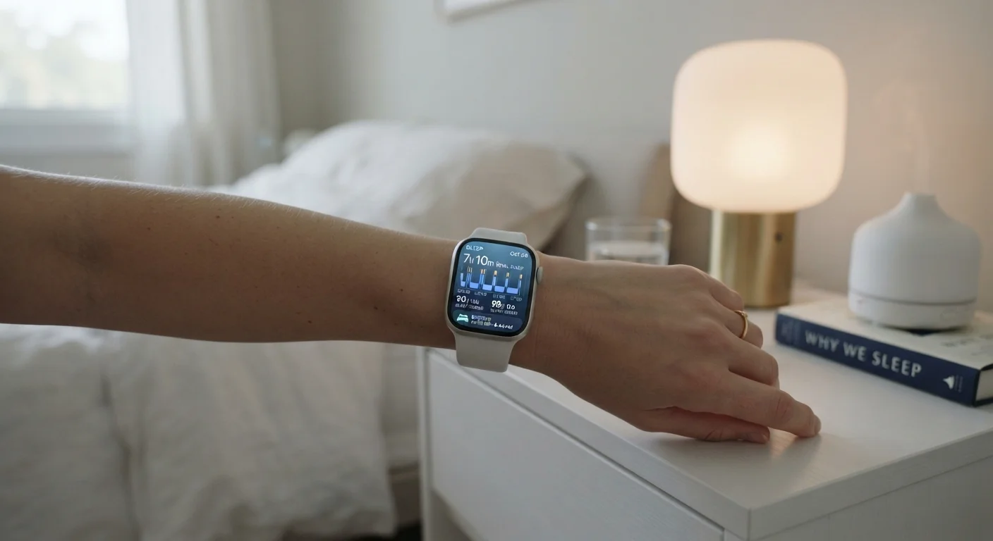
Consumer wearables and smartphone apps cannot replicate this level of detail. As explored in our article on what sleep trackers can and cannot tell you, wearables infer sleep stages from movement and heart rate rather than measuring brain waves directly.
What the Results Can Reveal — and What Comes Next
After the study, a credentialed sleep technologist scores the recording according to standardized AASM criteria, then a sleep medicine physician reviews the full report. The results may confirm a suspected disorder, rule one out, or surface an unexpected finding.
Common diagnoses that emerge from polysomnography include:
- Obstructive sleep apnea (OSA) — diagnosed by AHI severity (mild: 5–14 events/hour; moderate: 15–29; severe: 30 or more).
- REM sleep behavior disorder (RBD) — identified by abnormal muscle activity during REM and history of acting out dreams.
- Periodic limb movement disorder (PLMD) — confirmed by frequent, rhythmic leg movements during sleep.
- Narcolepsy — often requires an additional daytime test (the Multiple Sleep Latency Test, or MSLT) conducted the morning after the overnight study.
A negative or inconclusive result is also meaningful — it can redirect the clinical workup toward other causes of fatigue or poor sleep. Building sustainable sleep habits alongside any medical treatment is equally important; our overview of sleep hygiene principles covers the behavioral side of sleep health.
This article is for general informational and educational purposes only and does not constitute medical advice. If you are experiencing symptoms that concern you, consult a qualified healthcare professional for evaluation and guidance.